

Large Particle Detection (LPD)
- Simultaneous Submicron and Subvisible Monitoring
- Low Concentration Sensitivity
- Multiple measurements per minute
- Non-invasive and In-Flow
Advanced Non-Invasive Detection of Rare Subvisible Aggregates
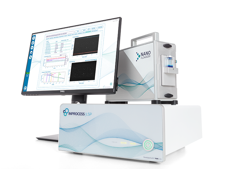
Large Particle Detection (LPD) is a powerful imaging technology integrated into the NanoFlowSizer, designed to detect and quantify rare large particles in nanoparticle suspensions and biopharmaceutical formulations.
Unlike conventional methods requiring separate instruments for different size ranges, the NanoFlowSizer combines Dynamic Light Scattering (PhaSR-DLS) and LPD imaging — enabling the detection of both submicron and subvisible aggregates in a single, non-invasive setup.
Using fast cross-sectional video imaging, LPD identifies oversized particles down to extremely low concentrations (<10³ particles/ml), complementing DLS measurements. This provides a complete, real-time understanding of particle populations, critical for safeguarding product quality and optimizing downstream processing (DSP).
Why Monitor Large Particles?
Oversized particles — whether originating from aggregation during manufacturing, storage, or transport — can compromise product stability, therapeutic efficacy, and safety.
In biopharmaceuticals, even low levels of subvisible particles (1–100 μm) can raise regulatory concerns and pose immunogenic risks.
Mechanical stresses, such as peristaltic pumping, are now proven to trigger aggregate formation over time. Studies using the NanoFlowSizer show a near-linear increase in both submicron and subvisible particle concentrations during prolonged circulation.
Detecting these aggregates early allows manufacturers to:
- Reduce filtration and chromatography clogging
- Increase product yields
- Maintain compliance with strict regulatory standards (e.g., USP<787>, USP<788>)
- Streamline quality control processes
Large Particle Detection
Key Advantages
- Direct detection of small quantities of larger particles in a suspension
- Supports Process Analytical Technology (PAT)
- Compatible with simultaneous SR-DLS measurements
- Significant time reduction in aggregation detection
Comprehensive and Real-Time Detection with LPD
Large Particle Detection (LPD) on the NanoFlowSizer offers a powerful and non-invasive method to detect, quantify, and monitor rare large particles in nanoparticle suspensions and biopharmaceutical formulations. Combining high-sensitivity imaging with dynamic light scattering, LPD provides comprehensive insights into submicron and subvisible particle populations in real time — all with a single instrument.
Your solution for:
- Detecting rare subvisible aggregates (>1 µm) at very low concentrations.
- Monitoring protein aggregation during manufacturing and transport.
- Real-time, non-invasive particle size characterization.
- Safeguarding biopharmaceutical product quality and stability.
- Meeting stringent regulatory requirements (e.g., USP<787>, USP<788>).
- Supporting process development and optimization in DSP.
What makes it unique:
- Dual Detection: Simultaneously monitors submicron and subvisible particles.
- Low Detection Limit: Detects down to <10³ particles/ml.
- Real-Time Insights: Immediate visibility into aggregation trends.
- Non-Invasive Analysis: No sample preparation or dilution required.
- Integrated Approach: Combines DLS and video imaging in a single measurement.
- Supports PAT Initiatives: Enables both inline and offline monitoring.
Application Example: Protein Aggregation under Mechanical Stress
In a recent study, BSA protein solutions subjected to peristaltic pumping were monitored over 20+ hours. Results demonstrated:
- Continuous growth of submicron aggregates (150–400 nm) detected by PhaSR-DLS.
- Increase in rare, large aggregates (>1 μm) detected by LPD imaging.
- Aggregate formation correlated directly with exposure time to mechanical stress.
- Both aggregation types were tracked simultaneously with the NanoFlowSizer.
This shows the crucial role of LPD in understanding and mitigating mechanical stress effects during critical DSP stages.
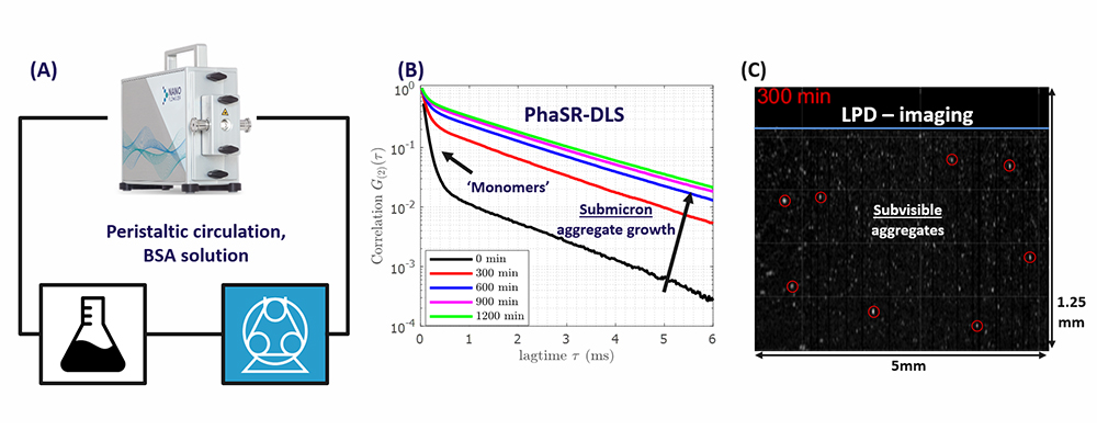
Figure 1. A) Peristaltic loop for circulating BSA solution, including the NanoFlowSizer (NFS) with a ¼ inch flow cell. B) PhaSR
measurements of DLS correlation functions of the BSA solution, after different circulation times. C) Snapshot of an LPD-movie,
after 5hr circulation. Encircled particles have an estimated size1 > 1𝜇𝑚. The blue line marks the glass wall of the flow cell.
Taking your process forward
More Applications
Related documents
Contact us:
Accessories & Software
Modules

LPD only requires a sample to be at an appropriate measuring position. This is ensured by our static modules: vials, flasks and customized.
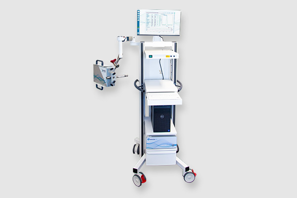
Trolley
Aliquam facilisis interdum mauris, venenatis eleifend libero dignissim in. Nullam vel arcu ligula. Sed ipsum ipsum,